For a surgeon performing an exploratory operation inside a patient’s abdomen, one of the greatest challenges is knowing what to look for, and where to look. Should you focus your attention in the near field, or far? How best to slowly advance your laparoscope a tiny distance at a time, to reduce the risk of missing a lesion or tumour just outside your field of view?
This enduring problem now has a solution thanks to the work of Keigo Iizuka, a professor in The Edward S. Rogers Sr. Department of Electrical & Computer Engineering. Professor Iizuka has designed and demonstrated the first omni-focus laparoscope, capable of focusing on microscopic images millimetres away and large objects 1.6 metres away simultaneously. This allows surgeons, already under mental and physical strain in the operating room, to see a much greater depth of field in one view without ever touching a focus dial, especially if dealing with a large lesion.

“Imagine you’re watching a concert on television,” Professor Iizuka says. “The singer might be in focus, but the orchestra isn’t. The omni-focus camera lets you see both the singer and orchestra in focus at the same time.”
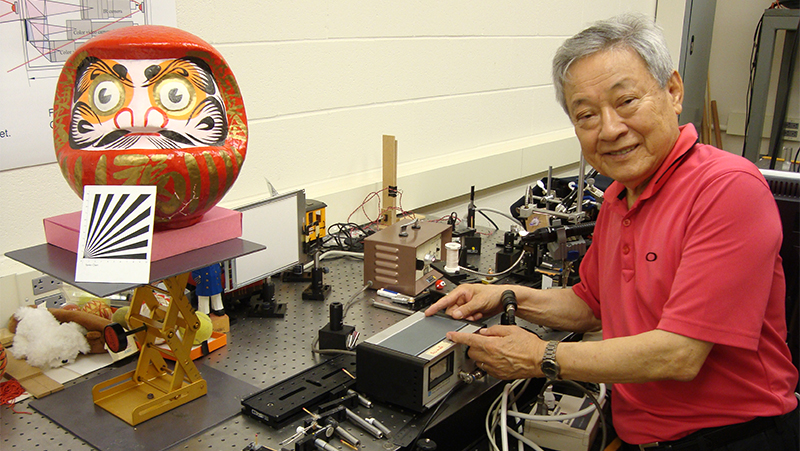
In demonstrating the system, Professor Iizuka shows the miniscule opening of a syringe on a fingerprint right in front of the laparoscope’s tip, and a large Japanese Daruma doll across the room, both in crystal-clear focus in a single image. He recently received an international patent for the system.
Professor Iizuka’s omni-focus device provides great advantages over even the latest model “chip in tip” laparoscope which typically allows a depth of focus of 10 centimetres. Autofocus means that a finite number of spots on the image are in focus at one time, while omni-focus means all points in the image are in focus at once, even over a much greater depth of field.
The experimental set-up uses a laparoscope 0.64 centimetres in diameter and 30 centimetres long, but could easily be modified to fit standard incision diameters of 0.5, 1 or 1.5 centimetres. One of the system’s main advantages is its simplicity: only the tiny laparoscope, with two light rings built along the shaft, travels inside the patient; the majority of the imaging and computing hardware is housed outside the body.
The omni-focus laparoscope is comprised of an array of infrared and colour video cameras aimed at the same point but focused at different distances, and a laparoscopic profilometer subsystem to measure the various distances captured in the field of view. A pixel switching subsystem selects pixels from the colour video cameras according to the distance measured by the profilometer, and compiles them into a single omni-focus image which is displayed on a screen.
The path of light through the lens relay is very similar to a low-power microscope, creating an opportunity for high-resolution magnification in vivo with only minor tweaks to the system. This could allow the omni-focus laparoscope to show biological features on the scale of bacteria, which range in size from four to 10 micrometers, or blood cells, at four to five micrometers in size.
Professor Iizuka hopes his next steps will be combining omni-focus capability with the magnifying power of conventional microscopes. Microscopes have thus far not been very useful in looking inside live patients because of their shallow depth of field—live objects quickly move out of focus. The omni-focus laparoscope’s much longer depth of field could eliminate this issue, completely changing the way diagnostic surgery is performed.
More information:
Marit Mitchell
Senior Communications Officer
The Edward S. Rogers Sr. Department of Electrical & Computer Engineering
416-978-7997; marit.mitchell@utoronto.ca